
 +31 85 483 31 03
Home
KneeReviver
For Healthcare Professionals
For Patients
Find a hospital
About us
Contact


≡
Discover Knee Joint Distraction
Osteoarthritis (OA) is the 11th leading cause of years lived with disability, according to the World Health Organization. Its prevalence is increasing markedly among adults under 55, indicating a growing need for effective joint management. In parallel, total knee arthroplasty (TKA) is increasingly performed in patients aged 50–65, a group with a substantial risk of revision. Younger adults now represent a significant proportion of TKA recipients, reinforcing the need to consider joint-preserving treatment strategies at an earlier stage.
Findings from research over the last year continue to underscore the challenges posed by OA, which continues to rise in prevalence, with more than 50% of the cases occurring in individuals younger than 55 years. OA prevention and early intervention remain thus critical to reduce the OA burden. (Dell’Isola, Andrea et al.)

Evidence shows that revision risk after total knee arthroplasty is not confined to patients under 50. Perdisa et al. report that individuals aged 50–65 represent the fastest growing TKA group and have a two fold higher revision risk than older adults, raising concerns about durability and long term suitability in this active population [Perdisa et al.].

Bayliss et al. report that total knee replacement patients younger than 60 years have a lifetime revision risk of 35 percent. Revisions occur most often within five years, with men and younger patients showing higher susceptibility. Mean time to revision ranges from 4.55 years at ages 50–59 to 3.57 years in older adults.

These developments highlight the growing pressure on knee care. With more than one third of all total knee arthroplasties performed in patients younger than 65 years, and a comparable 35 percent lifetime revision risk in this group, younger adults account for a substantial share of future revision procedures and long-term clinical burden.

Many orthopaedic surgeons regularly see younger, active patients who have exhausted conservative treatment but are not yet ideal candidates for knee replacement.
Complete the Treatment Gap Assessment to discover whether you are already treating patients who may benefit from Knee Joint Distraction.
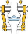
The principle behind Knee Joint Distraction is straightforward.
Create space.
Temporary distraction gently separates the joint surfaces.
Reduce stress.
Lower contact pressure temporarily unloads the joint.
Support natural repair.
By combining temporary unloading with continued weight-bearing, the treatment creates conditions that may support the knee’s natural repair processes.
For selected patients, this approach has been shown to reduce pain, improve function and delay knee replacement.




KneeReviver® is the only CE-certified medical device developed specifically for Knee Joint Distraction.
Unlike external fixation systems adapted from trauma surgery, KneeReviver® was engineered around the specific requirements of Knee Joint Distraction therapy.
Every aspect of the system supports predictable treatment, procedural efficiency and patient comfort.

KneeReviver was developed with one purpose: to perform Knee Joint Distraction safely, reproducibly and efficiently.

Assoc. Prof. Dr. Simon Mastbergen
Dr. Roel Custers, MD, PhD
Dr. Tim Boymans, MD, PhD
Prof. Dr. Med. Kaywan Izadpanah
Prof. Dr. Krik Heusdens, Mark Larkin, PhD,
Prof. Dr. Med. Christian Weber, Dr. med. Urs Hefti
Anzillotti G, Pavone S, Queirazza P, Conte P, Saccia F, Minelli M, Petrov P, Kon E, Di Matteo B. Cartilage. 2026 Jan 30. doi: 10.1177/19476035251409412
Objective: To bring together the available clinical, structural, biomarker and safety data on knee joint distraction (KJD) and implantable medial-compartment shock absorbers (ISA) for tibiofemoral knee osteoarthritis, and to compare these outcomes with arthroplasty, high tibial osteotomy, orthoses and non-operative care.
Design: A PRISMA-based systematic review of four major databases up to October 2025, identifying peer-reviewed clinical studies on KJD or ISA. Seventeen studies covering roughly 400 patients (13 on KJD, 4 on ISA) met the inclusion criteria.
Results: KJD produced clinically meaningful gains in pain and function (WOMAC/KOOS, VAS) within one to two years, with an estimated 75–85% of patients avoiding arthroplasty at five to nine years in selected series. These improvements were paired with increases in radiographic joint space width and MRI cartilage thickness in the most affected compartment.
Conclusions: Across small, heterogeneous cohorts of low-to-moderate certainty, both KJD and ISA appear capable of delivering meaningful short- to medium-term joint-preserving benefit.
Hamilton TW, Lineham B, Stocken DD, Pandit H; KARDS Study Group. Bone & Joint Open. 2025 Aug 6;6(8):886-893. doi: 10.1302/2633-1462.68.BJO-2024-0120.R2
Objective: To evaluate the clinical and cost-effectiveness of knee joint distraction (KJD) compared with knee arthroplasty in patients aged 65 or younger with symptomatic osteoarthritis unresponsive to non-surgical treatment (the KARDS trial, UK).
Design: Phase III, multicentre, pragmatic, randomized non-inferiority trial. Participants were randomized to static, linear 5 mm KJD (external fixator, 6 weeks) or total knee arthroplasty. The primary outcome was the KOOS pain subscale at 12 months. The trial closed early due to suspension of elective surgery during the COVID-19 pandemic.
Results: Only 24 participants were randomized (11 KJD, 13 arthroplasty). Median KOOS pain scores improved in both groups at 12 months (KJD: 38.9 to 55.6; arthroplasty: 30.6 to 75.0). The small sample size did not allow meaningful comparison between groups. Two KJD patients had pin site infections; one sustained a fracture after frame removal.
Conclusions: KJD appears to be associated with improved pain and function compared with baseline. Its clinical and cost-effectiveness relative to arthroplasty remains uncertain given the limited sample, though results are consistent with earlier studies where KJD performed well.
Lineham B, Joumah A, Hamilton T, Wijayathunga N, Pandit H.
Cartilage. 2025 Aug 4. doi: 10.1177/19476035251357836 — CC BY 4.0
Aims: Knee osteoarthritis (OA) is a significant source of morbidity and socioeconomic burden, exacerbated by aging populations and rising body mass index. Total Knee Replacement (TKR) is effective but may result in dissatisfaction or revision, particularly in young patients. Knee Joint Distraction (KJD) offers a joint-preserving alternative that may delay or avoid replacement. This study assessed cartilage morphology changes using magnetic resonance imaging (MRI) of patients up to 1-year post-KJD in patients from a randomized controlled trial (RCT). The primary aim was to evaluate cartilage volumes at 12 months post-KJD. Secondary aims were to evaluate additional MRI parameters for cartilage morphology and compare the MRI parameters with Patient-Reported Outcome Measure (PROM) scores at 3 and 12 months.
Conclusion: This study demonstrates improvements in cartilage MRI morphological parameters at 3 and 12 months following the emerging joint-sparing technique of KJD. Therefore, KJD has the potential to be considered as a surgical option for young patients with severe knee OA.
Vandenrijt J, Michielsen J, Verhaegen J, Van Dyck P, Heusdens CHW.
Journal of Experimental Orthopaedics. 2025 Oct 9. doi: 10.1002/jeo2.70459 — CC BY 4.0
Purpose: Knee joint distraction (KJD) offers a joint preserving alternative for relatively young patients with severe knee osteoarthritis (OA). The aim of this study was to assess the clinical and radiographic success of KJD in patients with severe OA.
Methods: This prospective case series comprised 26 consecutive patients undergoing KJD due to severe knee OA. Clinical and radiographic outcomes were collected at baseline and at 1- and 2-year follow-up. Complication, reoperation and conversion rates to a primary knee arthroplasty were collected. The patient-reported outcome measures (PROMs) were the Knee Injury and Osteoarthritis Outcome Score (KOOS), the 36-item Short Form Health Survey (SF-36) and the Visual Analogue Scale (VAS) for pain. The radiographs were analysed using an artificial intelligence (AI) based software package to extract compartmental joint space width (JSW), joint space area (JSA) and compartmental imbalance.
Conclusion: This case series demonstrates that KJD provides symptomatic relief amongst young patients with knee OA after failed conservative treatment. A significant increase in the JSW and JSA was observed, which may indicate cartilage regeneration. KJD could be an alternative joint-preserving intervention, with potential to postpone primary knee arthroplasty for young patients with knee OA.
Steijns JSJJ, Green D, Peeters LCW, et al. Connective Tissue Research. 2024;65(6):486-496. doi: 10.1080/03008207.2024.2440716
Purpose: Knee joint distraction has cartilage-regenerating potential, but the biomolecular makeup of the regenerated tissue in humans has rarely been studied. This case study offered a rare opportunity to characterize that tissue in a patient who underwent bilateral distraction and later required knee replacement in both knees.
Methods: Distraction was performed with an external fixation frame, with total knee arthroplasty following years later. Imaging tracked the joint before, during, and after each intervention. Cartilage biopsies taken at the time of joint replacement were processed for tissue sectioning, histochemical staining, and tandem mass-spectrometry proteomic analysis to compare regenerated and native cartilage.
Results: Both knees showed joint-space improvement following distraction. Regenerated cartilage appeared white and irregular, in contrast to the smooth, yellow native cartilage. Native cartilage stained more strongly for Safranin-O, and its collagen structure differed from the regenerated tissue. Despite these visual and histological differences, proteomic analysis found no major difference in the abundance of extracellular matrix proteins between the two tissue types. Bioinformatic analysis pointed to enrichment of ribosomal proteins in regenerated cartilage and of RNA Polymerase II transcription termination pathways in native cartilage.
Conclusion: Regenerated cartilage differs histologically from native cartilage — fewer glycosaminoglycans and a less organized collagen structure — but shows no major difference at the protein-abundance level. This points to protein translation regulation as a possible mechanism underlying the regenerative response.
Jansen MP, Salzlechner C, Barnes E, et al. Rheumatology (Oxford). 2023;62(8):2789-2796. doi: 10.1093/rheumatology/keac723
Objectives: Knee joint distraction (KJD) has previously been linked to clinical and structural improvement and to changes in synovial fluid markers. This study used an automated, AI-based measurement method to assess radiographic change after KJD and relate it to clinical outcome and synovial fluid markers.
Methods: Twenty knee osteoarthritis patients treated with KJD in regular care had radiographs and WOMAC scores collected before treatment and roughly one year afterward. Synovial fluid was sampled before, during, and after treatment and assessed by immunoassay. Radiographs were analyzed for compartmental joint space width, Kellgren-Lawrence grade, joint space narrowing, and osteophyte/sclerosis scores, separately for the most- and least-affected compartments.
Results: Sixteen patients had evaluable data. Joint space width, K-L grade, and joint space narrowing improved in roughly half of patients, reaching significance only for joint space width in the most affected compartment. That improvement correlated positively with reduced WOMAC pain. Larger increases in the marker MCP-1 and smaller increases in TGFβ-1 were both linked to greater joint space width improvement, and MCP-1 changes also correlated with pain reduction.
Conclusion: Automated radiographic analysis confirmed improved joint structure in most patients after KJD in regular clinical practice. The increase in joint space width in the most affected compartment was tied to synovial biomarker changes and to the pain relief patients experienced.
Struik T, Mastbergen SC, Brouwer RW, et al. RMD Open. 2023;9(2):e003074. doi: 10.1136/rmdopen-2023-003074
Objective: Knee distraction reliably postpones arthroplasty for years in end-stage osteoarthritis, but earlier studies relied on devices intended for other purposes, patient-personalized builds, or custom-made frames. This study is the first to evaluate a device purpose-built for knee distraction.
Design: Sixty-five patients aged 65 or younger with end-stage knee osteoarthritis and an arthroplasty indication underwent knee distraction. Questionnaires and knee radiographs were collected before treatment and at one and two years afterward, alongside records of adverse events and self-reported pain medication use.
Results: Forty-nine patients completed the two-year follow-up; one discontinued treatment early, three received arthroplasty in year one and four more in year two, and eight were lost to follow-up in the second year. Total WOMAC score improved by a clinically relevant margin at both one and two years (+26 and +24 points respectively, all subscales p<0.001), as did minimum radiographic joint space width (+0.5mm at one year, +0.4mm at two years) and the SF-36 physical component score (+10 points). Pin tract infection was the most common adverse event, affecting 66% of patients, successfully managed with oral antibiotics in 88% of cases; two patients needed hospitalization. Eight patients had device-related complications, none of which affected the two-year outcomes. Pain medication use nearly halved from 42% of patients at baseline to 23% at one year and 29% at two years.
Conclusions: A general-purpose device adapted for knee distraction, now replaced by one purpose-built for the procedure, produced significant clinical and structural improvement over two years, despite a meaningful rate of adverse events.
Jansen MP, Mastbergen SC, MacKay JW, Turmezei TD, Lafeber FPJG. Rheumatology (Oxford). 2022;61(3):974-982. doi: 10.1093/rheumatology/keab456
Objectives: Knee joint distraction (KJD) has previously shown long-term clinical improvement and short-term cartilage restoration in younger osteoarthritis patients. This study evaluated MRI-measured cartilage thickness up to 10 years post-treatment using a 3D surface-based method.
Methods: Twenty end-stage knee OA patients treated with KJD underwent MRI scans before treatment and at 1, 2, 5, 7, and 10 years afterward. Cartilage in the tibia and femur was segmented and mapped to a canonical surface for whole-joint statistical analysis, with compartmental thickness changes tracked over time using repeated-measures analysis.
Results: Cartilage thickness in the most-affected weight-bearing region was significantly greater than baseline at one and two years, gradually thinning after five years but remaining elevated at the 10-year mark. The least-affected compartment also showed significant long-term thickness gains. Male sex and more severe baseline osteoarthritis were weakly associated with a shorter-term benefit. Both tibia and femur showed significant short- and long-term thickness increases in the most-affected compartment.
Conclusion: KJD produces significant cartilage regeneration that is measurable on MRI both in the short term and up to 10 years after treatment.
Jansen MP, Struik T, Jaspers J, Mastbergen SC, Custers RJ. Journal of Cartilage Joint Preservation. 2021;1(2):100007. doi: 10.1016/j.jcjp.2021.100007
Introduction: Knee joint distraction (KJD) treats severe knee osteoarthritis at a relatively young age, but until recently this procedure was performed with devices designed for other purposes. Working with patients, clinicians, and medical device experts, a dedicated distraction (DD) device built specifically for KJD was developed.
Objectives: To compare user-friendliness between the new dedicated device and a previously used concept distraction (CD) device.
Methods: Patients were treated with one of the two devices (n=22 each). Intervention duration and complications were recorded, and after treatment patients completed a questionnaire covering practical difficulties around clothing, sleeping, pin care, daily activities, mobility, and complications.
Results: Average intervention time was 56 minutes with the CD device versus 44 minutes with the DD device (p<.001). Pin tract infection was the most common complication in both groups (73% CD vs. 55% DD; p=.210, not statistically significant). Of 34 patients who completed the questionnaire (16 CD, 18 DD), the DD device scored better on 6 of 25 questions (all p<.05), with no significant difference on the remaining items.
Conclusions: The dedicated device reduces surgery time and improves user-friendliness compared with the earlier concept device, supporting broader implementation of KJD treatment in regular clinical care.
Jansen MP, Van der Weiden GS, Van Roermund PM, Custers RJ, Mastbergen SC, Lafeber FP. Osteoarthritis and Cartilage. 2018;26(12):1604-1608. doi: 10.1016/j.joca.2018.08.004
Objective: Knee joint distraction (KJD) is a joint-preserving procedure for severe osteoarthritis that delivers clinical and structural improvement and delays total knee arthroplasty (TKA). This study assessed nine-year outcomes and identified which patient characteristics predict long-term treatment success.
Design: Twenty patients under 60 with severe tibiofemoral osteoarthritis who were candidates for TKA were treated with KJD. Questionnaires, radiographs, and MRI tracked outcomes over time, with “failure” defined as later receiving TKA.
Results: Nine-year survival (avoiding TKA) was 48% overall — 72% for men versus 14% for women, and 73% for patients whose joint space width increased by more than 0.5mm in the first year versus 0% for those below 0.05mm. Patients who avoided TKA still showed clear clinical improvement compared with baseline. Notably, even patients who eventually needed TKA years after KJD reported some lasting clinical benefit, though less pronounced. Patients who avoided TKA maintained a long-lasting increase in minimum joint space width, while those who didn’t avoid it did not. First-year joint space width and MRI cartilage thickness increase both predicted nine-year survival, as did male sex.
Conclusions: KJD shows long-lasting clinical and structural benefit. Male sex is associated with a substantially greater chance of long-term success, and how much cartilage repair activity occurs early on appears to be an important predictor of that success.
Jansen MP, Besselink NJ, van Heerwaarden RJ, et al. Cartilage. 2021;12(2):181-191. doi: 10.1177/1947603519828432
Objective: Knee joint distraction (KJD) and high tibial osteotomy (HTO) are both joint-preserving alternatives that postpone total knee arthroplasty (TKA) in younger osteoarthritis patients. This study reports two-year outcomes from two non-inferiority trials: KJD versus TKA, and KJD versus HTO.
Design: Patients eligible for TKA were randomized to KJD (n=20) or TKA (n=40). Separately, patients with medial compartment disease being considered for HTO were randomized to KJD (n=23) or HTO (n=46). Patient-reported outcomes were tracked over two years, radiographic joint space width was measured annually, and serum/urine collagen markers were assessed in the KJD groups.
Results: Of 114 patients available for two-year analysis, total WOMAC score improved significantly in every group (KJD-vs-TKA arm: +30.4 points for KJD, +42.4 for TKA; KJD-vs-HTO arm: +21.6 for KJD, +29.2 for HTO; all p<0.05), as did minimum joint space width (increases of 0.6–0.9mm across KJD and HTO groups, all p<0.05). Collagen type-II synthesis increased significantly at two years post-KJD. Half of KJD patients experienced pin tract infections, successfully treated with oral antibiotics.
Conclusions: KJD produced sustained clinical benefit and cartilage thickness increase over two years, and was clinically non-inferior to both HTO and TKA on the primary outcome.
van der Woude JAD, Wiegant K, van Roermund PM, et al. Cartilage. 2017;8(3):263-271. doi: 10.1177/1947603516665442
Objective: Total knee arthroplasty (TKA) may become unavoidable in end-stage knee osteoarthritis, but at a relatively young age this carries a real risk of future revision surgery. Joint-preserving alternatives such as knee joint distraction (KJD) are therefore preferred where possible. This paper presents five-year KJD follow-up data.
Design: Twenty patients under 60 with conservative-therapy-resistant tibiofemoral osteoarthritis who were TKA candidates were treated with KJD. Clinical evaluation used questionnaires, and cartilage thickness change was quantified on radiographs and MRI. Five-year changes were compared against the natural progression of osteoarthritis using Osteoarthritis Initiative reference data.
Results: At five years, patients still reported clinical improvement from baseline (WOMAC +21.1 points; pain VAS -27.6mm), and minimum radiographic joint space width in the most-affected compartment remained increased (+0.43mm). Mean joint space width and mean MRI cartilage thickness were no longer statistically different from baseline at this point. Regression analysis showed KJD was associated with significantly less progression in joint space width and cartilage thickness compared with the natural disease course.
Conclusions: KJD produces prolonged clinical benefit, likely explained by an initial surge of cartilage tissue repair that provides a lasting structural advantage relative to how the disease would otherwise have progressed.
Van der Woude JAD, Nair SC, Welsing PM, Castelein RM, Van Laar J, Lafeber FPJG.
PLoS One. 2016 May 12;11(5):e0155524.
Objective: In end-stage knee osteoarthritis the treatment of choice is total knee arthroplasty (TKA). An alternative treatment is knee joint distraction (KJD), suggested to postpone TKA. Several studies reported significant and prolonged clinical improvement of KJD. To make an appropriate decision regarding the position of this treatment, a cost-effectiveness and cost-utility analysis from healthcare perspective for different age and gender categories was performed.
Methods: A treatment strategy starting with TKA and a strategy starting with KJD for patients of different age and gender was simulated. To extrapolate outcomes to long-term health and economic outcomes a Markov (Health state) model was used. The number of surgeries, QALYs, and treatment costs per strategy were calculated. Costs-effectiveness is expressed using the cost-effectiveness plane and cost-effectiveness acceptability curves.
Results: Starting with KJD the number of knee replacing procedures could be reduced, most clearly in the younger age categories; especially revision surgery. This resulted in the KJD strategy being dominant (more effective with cost-savings) in about 80% of simulations (with only inferiority in about 1%) in these age categories when compared to TKA. At a willingness to pay of 20.000 Euro per QALY gained, the probability of starting with KJD to be cost-effective compared to starting with a TKA was already found to be over 75% for all age categories and over 90-95% for the younger age categories.
Conclusion: A treatment strategy starting with knee joint distraction for knee osteoarthritis has a large potential for being a cost-effective intervention, especially for the relatively young patient.
KneeReviver® is intended for selected patients with symptomatic knee osteoarthritis who may benefit from joint preservation. It is especially relevant for younger, active patients where total or partial knee replacement is considered less desirable because of implant lifespan and revision risk. Final patient selection remains the responsibility of the treating orthopaedic surgeon.
Knee joint distraction temporarily unloads the tibiofemoral joint by creating controlled separation between the joint surfaces. This aims to reduce mechanical stress and create conditions that support joint tissue repair and symptom improvement.
The surgical procedure is relatively short, often around 30 to 45 minutes. The KneeReviver® typically remains in place for approximately six weeks, after which the device and bone pins are removed during day-care treatment.
Long-term clinical data show that knee joint distraction can reduce pain, improve function and delay knee replacement in selected patients. The ArthroSave whitepaper highlights pooled long-term data from 132 patients across five studies, with a mean treatment survival of 7.2 years and a 48% survival rate at 10 years.
The pathway includes patient assessment, diagnosis, discussion of treatment options, pre-operative preparation, surgical placement of the KneeReviver®, post-operative distraction, daily monitoring, wound and pin-site care, physiotherapy, outpatient follow-up with X-ray, device removal after six to seven weeks, and continued rehabilitation.
Hospitals can contact ArthroSave for product availability, procedural support, training and implementation guidance. During initial procedures, ArthroSave can support the surgical team with product expertise and practical workflow knowledge.
Every patient pathway is different. ArthroSave can support your team with product information, procedural guidance and implementation questions. You can also contact us to discuss KneeReviver® availability for your hospital.
Whether you’re evaluating the treatment, preparing your first procedures or expanding an existing programme, ArthroSave supports implementation through clinical training, procedural guidance and ongoing support.